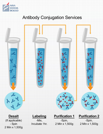
Antibody Conjugation Services
Conjugate many of Aviva's antibodies by request with these label options for $45:
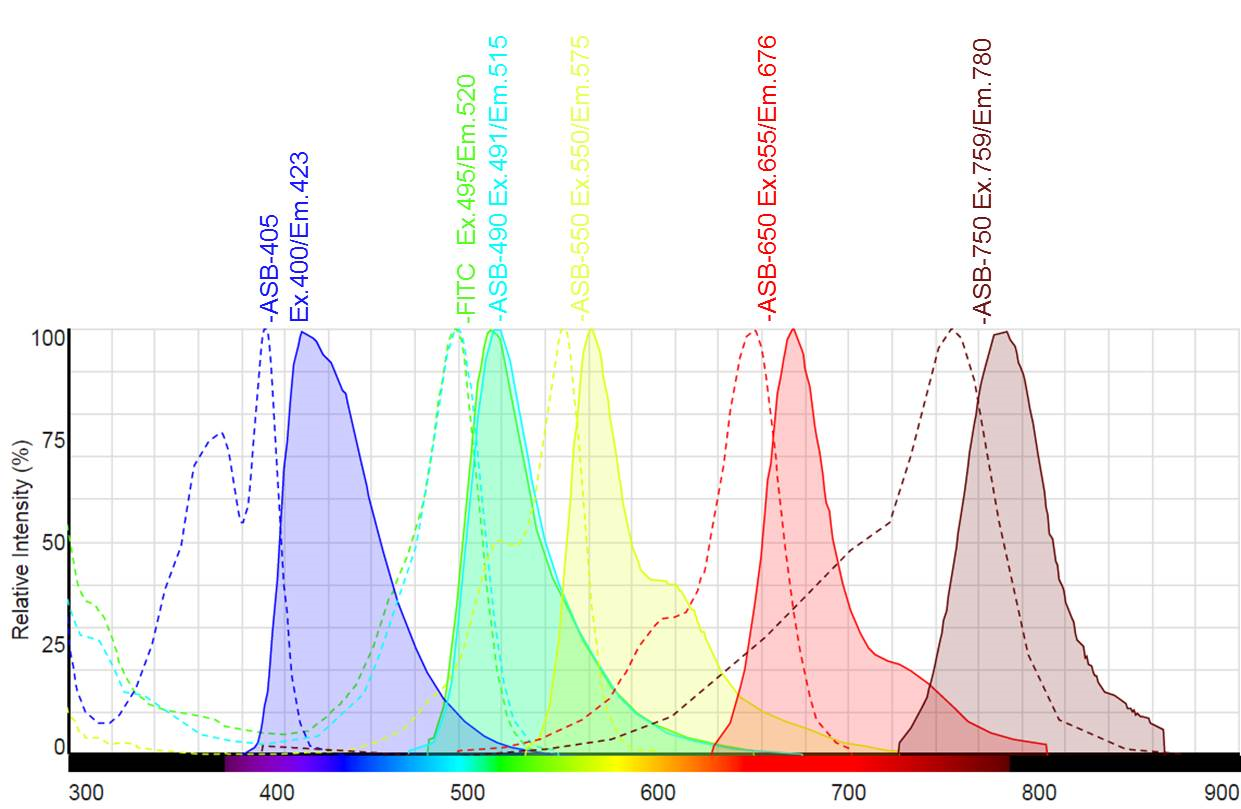
| Conjugate | Excitation | Emission |
|---|---|---|
| FITC (FAM) Fluor | 495 nm | 520 nm |
| ASB-405 Fluor | 400 nm | 423 nm |
| ASB-490 Fluor | 491 nm | 515 nm |
| ASB-550 Fluor | 550 nm | 575 nm |
| ASB-650 Fluor | 655 nm | 676 nm |
| ASB-750 Fluor | 759 nm | 780 nm |
| Biotin-Peg4 | - | - |
| Horseradish Peroxidase | - | - |
The reduced complexity of using Aviva's direct conjugate primaries provides numerous process and ease-of-use advantages over traditional two-step detection methods:
- Shorter assay procedure times - higher throughput
- Simpler assay optimization - less development time
- Reduced non-specific interactions - a single affinity reagent opposed to two
- Lower biological assay reagent costs
Simply use the drop-down menu found on antibody data sheets to specify which label is required along with the antibody order:

Technology
Aviva utilizes a proprietary protein conjugation technology to conveniently produce directly labeled primary antibodies for immunohistochemistry, immunofluorescence and other applications. Side-by-side comparison shows Aviva's conjugated antibodies providing superior results to comparable labeling methods in IHC. High signal intensity and specificity indicates the conjugation does not interfere with the antibody binding domains. Absence of non-specific background is attributable to highly effective final purification, lacking with other conjugation methods employing quenchers to chelate unbound label.
Example 1: IHC on human lung tissue using an NFATC4 antibody labeled with FITC using three different vendor's conjugation methods.
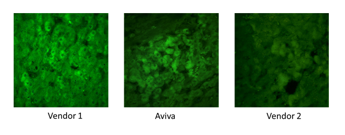
Conclusion - Vendor 1 and Aviva's Conjugated Antibodies produce uniform cytoplasmic staining while Vendor 2 conjugated antibody staining is noticeably weaker. Over-saturation causes greatly reduced cellular resolution in Vendor 1's stain when compared to that of the Aviva's conjugated stain.
Example 2: IHC of human liver using an ADCK2 Antibody labeled with FITC using three different vendor's conjugation methods.
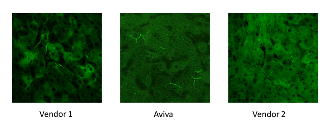
Conclusion - Antibody conjugated using Vendor 1 method produces extensive non-specific cytoplasmic staining, as well as non-optimal overstaining intensity in the bile canaliculi. Aviva's Conjugated Antibodies produces strong specific signal in canaliculi and cytoplasmic signal is absent as expected. Antibody labeled using Vendor 2 method does not detect canaliculi, and exhibits Vendor 1's non-specific cytoplasmic staining with weaker intensity.
Multiplexing IHC with Aviva Conjugated Primaries
Information pertaining to several co-localized biomarkers or organelles are typically required when performing IHC on a single tissue sample. Multiplexing detection of several targets provides numerous benefits.
- Increased throughput
- Improved consistency of relative measurements
- Reduced staining/assay time and costs compared to two-step detection
- Simplified assay development time compared to more complex two-step detection
- Specifically designed fluorescent dyes in regards to:
- Optimized discrete Excitation/Emission wavelengths for multiplexing
- Bright emission providing high signal intensity and low background for optimal sensitivity
- Greater multiplexing options compared to combinations limited by a species-affinity based two-step system
Directly labeled primary antibodies facilitate the easiest method for multiplex IHC applications. Combining Aviva's extensive antibody catalogue along with flexible conjugation options provides the ability to perform multiplexed detection of greater than 4 biomarkers, including DAPI, on a single tissue specimen.
Figure 1. Emission wavelength spectra of the Aviva dye conjugates are optimized for multiplexed applications.
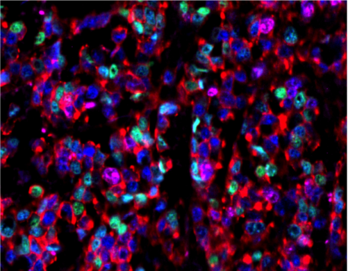
Example 1: Multiplexed fluorescent staining of a single triple-positive breast cancer tissue using HER2 / ASB-405 (red), Estrogen Receptor / ASB-490 (ER, blue), Progesterone Receptor ASB-550 (green) rabbit polyclonal antibodies and Ki-67 (magenta) using mouse monoclonal antibody.
Antibody Conjugation Kits
Aviva also provides convenient kits including all required reagents along with easy to use protocols for the conjugation of 100 ug antibody. The rapid conjugation protocol takes approximately 1 hour with around 15 minutes hands on time.
Fluor
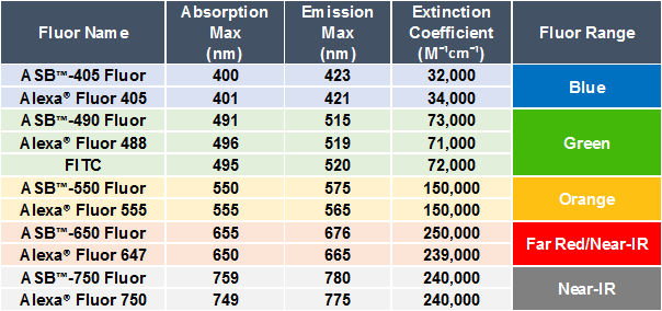
Aviva's proprietary ASB® Flours are a direct substitute for similar organic fluorophores such as Alexa™ Fluors or dyes such as FITC.
- Aviva ASB™ Fluors can be imaged on existing instrumentation for Alexa® Fluor or FITC Dyes.
- Respective adsorption and emission spectra are identical between fluors.
- Extinction coefficients show brightness and degradation rates are the same.
- Functional staining shows identical results between fluors.
Figure 1. Table comparing the excitation (nm), emission (nm) spectra and extinction coefficients (M¯¹ cm¯¹) between Aviva's organic ASB™ Fluors and Alexa® Fluor. All parameters are virtually identical.
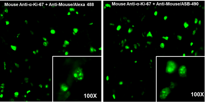
Example 1. Comparison of IHC staining intensity and specificity between Alexa® Fluor 488 and ASB® Fluor 490. Human breast cancer tissue was first stained with a monoclonal mouse anti-α-Ki-67 antibody. Stained tissues were then detected using a goat anti-mouse IgG antibody conjugated with either Alexa® Fluor 488 and ASB® Fluor 490. Excitation at 496 nm and image capture at 519 nm produce identical results between fluors.
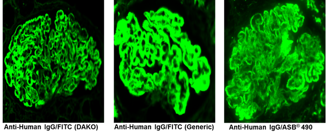
Example 2. Comparison of staining intensity and specificity between FITC dyes and Aviva ASB® 490. Kidney tissue samples from subjects exhibiting membranous glomerulonephritis were stained with anti-Human IgG antibodies conjugated with either DAKO. Inc. FITC, a generic FITC or Aviva ASB® 490. Excitation at 496 nm and image capture at 519 nm produce identical results between DAKO and Aviva stains. The generic FITC exhibits some non-specific staining.